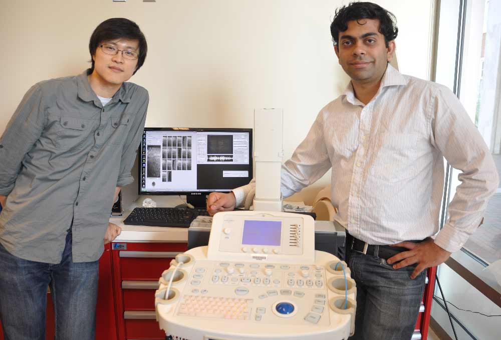
Dr. Nishikant Deshmukh (right) with his colleague and labmate Dr. Hyun Jae Kang with the altered ultrasound machine.
Dr. Nishikant Deshmukh (right) with his colleague and labmate Dr. Hyun Jae Kang with the altered ultrasound machine.
SAN FRANCISCO (Diya TV) – An Indian researcher at Johns Hopkins University has developed the world’s first five-dimensional ultrasound system, a breakthrough in medical science that will allow physicians to better detect and treat cancerous tumors.
Nishikant Deshmukh, who only just earned his doctoral degree in computer science from the prestigious institution, developed the technology as part of his Ph.D. thesis.
You can visit the Butterfly Network website if you want to find more info, but current ultrasound technology used by most of the world’s surgeons is predominantly two-dimensional, while some hospitals are fortunate enough to have three-dimensional devices. However, that technology does not update in real time and takes longer to generate images. This complicates matters for surgeons who seek to use the information while in the operating room.
Deshmukh’s technology will combine 3D ultrasound B-mode and the 3D ultrasound elastography volumetric data and make them available in real-time. Elastography is a medical imaging method that measures elastic properties of soft tissue and maps them as an image for diagnosing stiff regions such as cancer tumor. B-mode images are those that come across during a doctor’s sonography scan. Sonography, or diagnostic ultrasound, is a medical imaging technology where sound waves are used to produce images.
Deshmukh’s development has been termed as five-dimensional because of its ability to visualize and get the current combined data in real-time. The advanced imaging model that he developed can generate elastography using Graphic Processing Units at 60-70 frames per second, which enables combining elastography with real-time machine-generated B-mode images.
He presented the findings from his research for the first time to the public in 2015 at the Information Processing in Computer Assisted Interventions, a premier forum in the field of medical science. Deshmukh has also published the research, along with his advisors and colleagues at the Laboratory of Computational Science and Robotics at the Johns Hopkins University and the National Institutes of Health, in two journals, the International Journal of Computer Assisted Radiology and Surgery and PLOS ONE.
Deshmukh said the technology will be best used for early stage cancer detection, specifically for prostate and breast cancer.
“It will help a radiologist to determine whether the abnormally grown tissue is a potentially fatal tumor, or a more benign cyst,” he said.